Battle with creeping roots
- Dr. Vera Teh

- Feb 20, 2024
- 4 min read
Updated: Nov 30, 2024
Vascular lesions, encompassing a spectrum of abnormalities affecting blood vessels, can impact arteries, veins, and capillaries. While often harmless, many individuals seek ways to diminish their appearance for cosmetic or discomfort-related reasons. These lesions are attributed to aging, oxidation, and various factors that cause vascular dilation, damage, and changes in connective tissue.
Unveiling Vascular Lesion
Vascular lesions are broadly classified as congenital and acquired.
Congenital vascular lesions:
a. Hemangioma

Known as “strawberry mark”, is a benign vascular tumor derived from blood vessel cell types.
b. Port wine stain

Port wine stain (PWS) occurs in 0.3 to 0.5% of newborns initially as a light-pink macule or patch anywhere on the body.Over time, the color may deepen and become dark-red or violaceous with associated hypertrophy and nodularity.
Acquired vascular lesion:
a. Telangiectasia
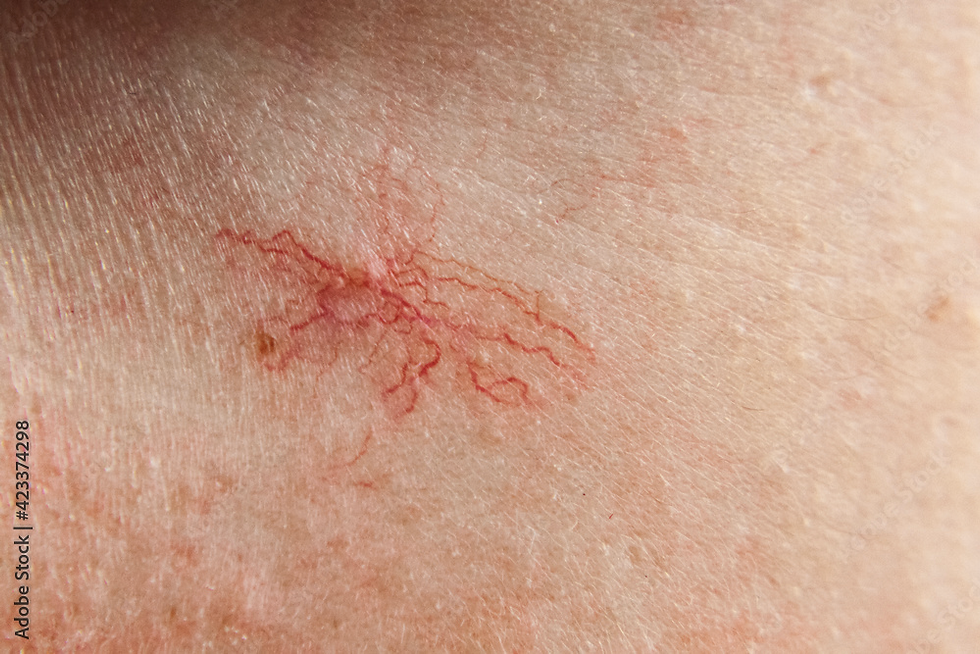
Telangiectasias are commonly occurring cutaneous vessels that typically measure 0.1 to 1.0 mm in diameter. Origins of these lesions aret dilated capillary, small venule or arteriole. They are also classified into groups, such as linear (or simple), arborizing, spider, and papular. The most commonly affected areas are the alar, dorsum and tip of the nose, as well as the ma- lar region. Significant sun exposure, hormonal factors, or genetic predisposition may all play a role in telangiectasia development. (Goldman, A. et al, 2021)
b. Rosacea associated telangiectasia

Rosacea is an acute skin condition that usually develops on the central areas of the face. Common symptoms include a profusion of inflamed papules and pustules; flushing of the face; erythema and telangiectasia.
c. Cherry angioma

Cherry angiomas are common, benign cutaneous vascular tumors, which commonly appear on trunk or extremities. They are most common on the trunk and proximal extremities and are often found in large numbers.
d. Venous lake

Venous lakes are lesions of dilated venous capillaries on the face, lips, and ears of older adult patients. The lesions bleed easily following minor trauma.
e. Reticular veins

They are dilated veins that are tortuous, protruding, bluish, and typically between 1 and 3 mm in diameter. They are typically found in periorbital and temporal region (Lee T.S et al, 2020).
f. Cutaneomucosal venous malformations

They present as multiple small, superficial lesions of various hues of blue that are easily compressible. Lesions involve skin and oral mucosa at the cervicofacial area and sometimes extremities.
Understanding the Mechanism of Laser
The goal of laser treatment is to induce vessel wall damage through destruction of hemoglobin while minimizing injury to adjacent structures. With longer pulse-widths or stuttered pulses, photothermal damage occurs. This causes slow heating of the vessel, intravascular coagulation, and collagen contraction. Clinically, one sees immediate blanching or subtle darkening of the vessel followed by subsequent erythema and edema. As a result, the blood supply to the affected area is maintained even as destroyed cells are flushed away by the body’s immune system.
It is worth mentioning that wavelength selection plays a pivotal role, ensuring effective treatment of deeper vascular lesions without causing adverse effects like dyspigmentation. Ultimately, the efficacy of laser treatment depends on various factors, including the target chromophore, vessel diameter, vessel depth, site of lesion, vascular flow rate, skin type, patient age, spot size availability, fluence, and the presence of epidermal cooling.

So Why Fotona SP Dynamis Nx Line?
The Fotona SP Dynamis Nx Line, equipped with a long-pulsed Nd:YAG system, offers a tailored approach (Kozarev , J., 2011; Vesnaver, A. et al, 2009). With precise depth control and variable pulse duration, it effectively addresses both superficial and deep vascular lesions (Lee T.S. et al, 2020). Its safety features, including optimal cooling systems, ensure patient comfort and safety during treatment.
Synergizing with Fotona StarWalker PQX
Combining treatments with Fotona StarWalker PQX enhances the therapeutic approach. This combination targets various skin issues like acne, pigmentation, melasma, rosacea, and facial veins by focusing on different skin layers, resulting in smoother, fairer, and firmer skin.
Differentiating Fotona SP Dynamis Nx Line from Traditional IPL
While intense pulsed light (IPL) treatments offer versatility in addressing both vascular and pigmented lesions, the Fotona SP Dynamis Nx Line stands out for its precision, adaptability, and safety measures, making it a preferred choice for vascular lesion treatment (Yepuri, V. et al, 2021).
Treatment Process and Aftercare
Typically, one to two treatment sessions, spaced 4 to 6 weeks apart, are found to be sufficient. Patients may notice improvements within weeks, with gradual enhancement over time. Post-treatment care involves measures to manage transient redness and swelling, such as hydrating masks and sunscreen application.
Ready to Embrace Flawless Skin?
Bid farewell to vascular lesions. Schedule a consultation with our experts at dream clinic today. Discover how the advanced Fotona SP Dynamis Nx Line can revitalize your skin and restore your confidence.

References
Goldman, A., Wollina, U., Machado, D., & Marinowic, D. (2021). LONG-PULSED ND:YAG LASER TO TREAT TELANGIECTASIA OF THE NOSE: A COMPREHENSIVE 5-YEAR SINGLE CENTER STUDY. GeorGian Medical News, 5.
Kozarev , J. (2011). Use of long pulse nd:YAG 1064nm laser for treatment of rosacea telangiectasia. https://www.laserandhealthacademy.com/media/objave/academy/priponke/5_use_of_long_pulse_nd_yag_1064_nm_laser_for_treatment_of_rosacea_telangiectatica.pdf
Lee, T. S., Kwek, J. W., & Ellis, D. A. (2020). Treatment of periocular and temporal reticular veins with 1064‐nm nd:YAG laser. Journal of Cosmetic Dermatology, 19(9), 2306–2312. https://doi.org/10.1111/jocd.13504
Vesnaver, A., & Dovšak, D. A. (2009). Treatment of large vascular lesions in the orofacial region with the nd:YAG laser. Journal of Cranio-Maxillofacial Surgery, 37(4), 191–195. https://doi.org/10.1016/j.jcms.2008.10.006
Yepuri, V., Patil, A. D., Fritz, K., Salavastru, C., Kroumpouzos, G., Nisticò, S. P., Piccolo, D., Sadek, A., Badawi, A., Kassir, M., Gold, M. H., Große-Büning, S., Grabbe, S., & Goldust, M. (2021). Light-based devices for the treatment of facial erythema and Telangiectasia. Dermatology and Therapy, 11(6), 1879–1887. https://doi.org/10.1007/s13555-021-00607-8


